Using Biotechnology to Detect and Treat Disease
Let
it is any disease, ranging from a fever (which is the symptom of many diseases)
to HIV, COVID-19, diagnosis at an early stage is very important for giving specific
treatment. Whenever I go to a doctor, he asks me things like stomach pain,
fever, body pain, headache, running nose, throat pain, etc., then, he checks
pulse, then he uses his stethoscope, he then gives all the possible
antibiotics! Yes, really, this happens in most cases. Then, if my problem is
not fixing up in a week, he'll ask me to take a blood test, this, that, and,
all!
Nothing wrong in this, because he
can't ask everyone coming to him to take a blood test at the very first sitting,
then, no one will visit him back saying he sucks out blood all the time! But,
it is okay to have a blood test after a week, but, not after a month!
Diagnosing a disease after it had reached a severe stage is comparatively less
significant than diagnosing at an early stage. So, how to diagnose?
Antigen-Antibody!
Antigen and Antibody interactions are mainly used for diagnosing most of the diseases. When you give your blood sample to your doctor, he gives it to his lab, where they check for the presence of the particular antigen in your blood. For example, if you have typhoid, surface proteins of the causative organism (Salmonella typhi) will act as antigen and when antibody (specific protein produced by your body against the antigen) is added to the infected blood sample antibody-antigen interaction occurs, which could be detected by various technique

Molecular Diagnosis: Antigen and Antibody interactions are mainly used for diagnosing most of the diseases. When you give your blood sample to your doctor, he gives it to his lab, where they check for the presence of the particular antigen in your blood. For example, if you have typhoid, surface proteins of the causative organism (Salmonella typhi) will act as antigen and when antibody (specific protein produced by your body against the antigen) is added to the infected blood sample antibody-antigen interaction occurs, which could be detected by various technique

In the last decade, the molecular diagnostics field has been growing rapidly. Tests or assays used in molecular diagnostics detected specific DNA or RNA sequences. Specific molecular probes and primers are designed for this purpose. Molecular diagnostics tests or

There are several methods available for diagnosing a disease, but, most of them are based on antigen-antibody interaction. Here I had listed a few methods of diagnosis, which I know:
 |
Possible methods for diagnosing a disease!
|
1) ELISA - Enzyme-Linked Immunosorbent Assay!
This method is based on antigen-antibody interaction.
Two antibodies are used for detecting the presence of a single antigen. But,
both the antibodies won't be directly reacting with the antigen, one antibody
(primary) will be reacting with the antigen, and another antibody (secondary
-which was raised against the primary antibody) will react further with the primary antibody used, producing color based on the enzyme-linked with it.
Generally done using 96 wells plate.

2. Dipstick colloidal dye immunoassay (DIA)- Lateral flow strips for detection of viruses
lateral
flow tests realized on nitrocellulose strips with immobilized antibodies
against the virus. A sample containing virus flow by
capillarity from the sample pad to bind test and control lines. In contrast, a
sample without target virus flows from the sample application pad and binds
only to the control line. The test combines the concepts of double antibody (Ab) sandwich ELISA, dot blotting, and colloidal particle-linked Abs to produce a dipstick test for multiple antigens (Ag) detections. Dipsticks prepared from Ab coated nitrocellulose membrane mounted on acetate strips served as the assay capture matrix. Abs absorbed to colored dye particles from a family of commercially available textile dyes (Dye/Ab reagent) served as Ag detecting reagents


3. Western Blot
This is also done for finding the presence of a specific protein (in diagnosis, the protein is antigen) in a given sample. This is done generally after doing SDS PAGE and transferring the protein separated in the gel to nitrocellulose membrane followed by a similar technique of adding primary and secondary antibody, which will show color if there is infection.
This is also done for finding the presence of a specific protein (in diagnosis, the protein is antigen) in a given sample. This is done generally after doing SDS PAGE and transferring the protein separated in the gel to nitrocellulose membrane followed by a similar technique of adding primary and secondary antibody, which will show color if there is infection.
4. PCR
PCR is used in the diagnosis of diseases that have become very common in laboratory practice. Its advantages (speed, sensitivity, specificity) are far more important than its drawbacks (risk of contamination, sensitivity to inhibitors, complexity, and cost) and several modifications to solve these problems have been performed with success. In general, PCR, with all its variants, is currently a basic tool in diagnosis, alone or preferentially in combination with other techniques
PCR is used in the diagnosis of diseases that have become very common in laboratory practice. Its advantages (speed, sensitivity, specificity) are far more important than its drawbacks (risk of contamination, sensitivity to inhibitors, complexity, and cost) and several modifications to solve these problems have been performed with success. In general, PCR, with all its variants, is currently a basic tool in diagnosis, alone or preferentially in combination with other techniques
Nested PCR
Sensitivity and specificity problems
associated with conventional PCR and RT-PCR can be reduced
by using nested PCR-based methods,
based on two consecutive rounds of amplification (Fig. 1). Usually, the products
of the first amplification are transferred to another tube before the nested
PCR is carried out using one or two internal primers (if one is used it is semi
nested or more than 1 primer is used then it is nested PCR )

Multiplex PCR
The simultaneous detection of two or
more DNA or/and RNA targets can be afforded by duplex or
multiplex PCR in a single reaction
with several specific primers included in the PCR cocktail. Multiplex PCR is
very useful in plant pathology because different bacteria or viruses frequently
infect a single crop or host. In multiplex assays where closely related
templates such as pathogen strains are distinguished by amplifying differing
sequence, primers for a sequence common to all templates provide a positive
control for amplification.
RT-PCR
The synthesis of DNA
from an RNA template, via reverse transcription, produces complementary DNA (cDNA). Reverse transcriptases (RTs) use an RNA
template and a short primer complementary to the 3' end of the RNA to direct
the synthesis of the first-strand cDNA,
which can be used directly as a template for the PCR. For some cDNA generated, it may be
necessary to remove the RNA complementary to the cDNA
prior to PCR amplification. This is particularly
applicable to PCR targets of 1 kb or larger. Add 2 units of RNase H to the cDNA generated and
incubate at 37oC for 20 minutes

Real Time qPCR
Two primers flank the sequence of interest and a third fluorescently labeled primer anneals between them. As the flanking primers extend, the labeled primer is released and fluorescence occurs The advantages of this method are that no post-reaction processing is required to detect and that it is quantitative. Generally SYBR Green dye,
Taqman or molecular beacon probes are used for Real-Time PCR. In SYBR Green the method, the fluorophore binds with the double-stranded DNA and produces
fluorescence, and hence as the amplification increases, fluorescence increases,
but the fluorophore has no specificity and hence, even if the amplified product
is not the product of your interest you would find an increase in fluorescence.
The TaqMan and molecular beacons are probes that bind which are labeled with
fluorescent dyes at one end and quencher at another end. When the fluorescent dye
and the quencher are at close proximity there won’t be any fluorescence but on
amplification, the fluorescent dye and the quencher get apart thus causing the increase in fluorescence. These are specific to the gene amplified and hence we
could monitor the amplification specifically online. Melting curve analysis is
generally done for the PCR products in the case of LUX and SYBR Green as the
fluorescence could also be due to the primer dimer.


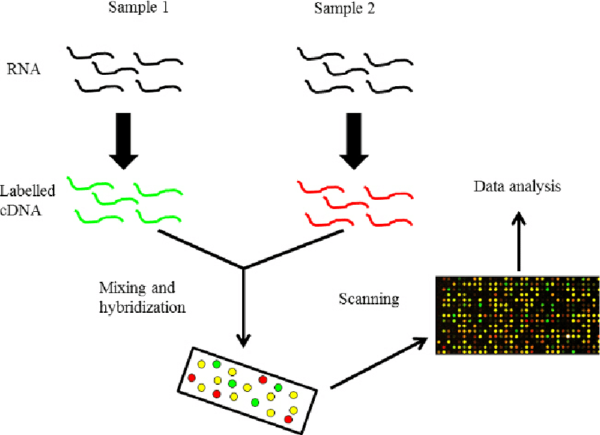
5. Microarray
Technology
Since the development of microarray
technology for gene expression studies, new approaches are
extending their application to the
detection of pathogens. Microarrays are generally composed of thousand of specific probes spotted onto a
solid surface (usually nylon or glass). Each probe is complementary to a specific DNA sequence provides a
signal that can be detected and analyzed. Although there is great potential for microarray technology in the
diagnosis of plant diseases, the practical development of this application is still in progress
6. Loop-mediated Isothermal Amplification (LAMP
It is a type of isothermal amplification that it is being increasingly used in the diagnostic field offering
sensitivity and economic costs. The method requires a set of four specifically designed primers that recognize six distinct sequences of the target and a DNA polymerase with strand displacement activity. The amplification products are stem-loop DNA structures with several inverted repeats of the target and cauliflower-like structures with multiple loops, yielding >500ug/ml. The LAMP reaction was enhanced by the addition of loop primers, reducing time and increasing sensitivity The amplification takes place at 60-65 C for 60 min. Although it was initially developed for DNA it can be adapted to amplify RNA (RT-LAMP).

I prefer to go with PCR than any other technique, but, when there is no thermocycler in your laboratory it's better to go with LAMP / ELISA! There are also other techniques like Biosensors, latex agglutination and microscopic observation of samples, but, the best the way is PCR!
Hope this helped you, any doubts? Then, mail me, happy to clear your doubts (if I can, as I'm not an expert )
- RUSHIKESH RAVINDRA TAHAKIK
tahakikpatil@gmail.com
M.Sc. Plant Biotechnology
Genetics and Plant Breeding,BHU Institute of Agril Sciences
Varanasi, UP India
M.Sc. Plant Biotechnology
Genetics and Plant Breeding,BHU Institute of Agril Sciences
Varanasi, UP India
Comments